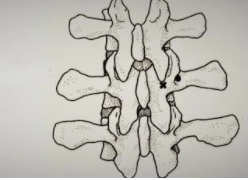
下颈椎前路椎弓根钉(Cervical anterior pedicle screw, CAPS)具有有效的稳定性、避免后路手术的优点[1-6],已有用于临床的报道 [7-10]。目前CAPS的植入采用的是椎弓根轴位透视方法,但仍然存在着脊髓、椎动脉、神经根损伤的风险[7-10]。随着术中CT和导航技术的发展,已有采用术中O-Arm 3D成像加导航应用于下颈椎后路椎弓根钉植入的报道[11]。本文采用同样的方法,首次报道1例应用于颈椎小关节骨折脱位前路椎弓根植入,并取得满意结果。
1.病例报道
1.1一般资料。男,52岁。因车祸伤后颈痛伴四肢左上肢麻木无力3天入院。查体:左上肢桡侧、中环指痛觉减退,左上肢伸肘、伸腕、握力肌力IV级,余四肢肌力V级,四肢腱反射正常。CT示颈6、7椎左侧小关节突骨折,颈6/7左侧小关节脱位。MRI示颈6/7椎间盘左侧椎间孔突出,脊髓无受压(图1)。诊断:颈6/7左侧小关节骨折脱位,颈7神经根损伤。
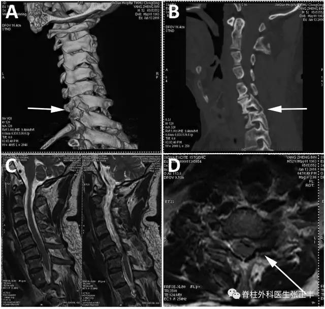
图1. (A,B)CT示颈6、7椎左侧小关节突骨折,颈6/7左侧小关节脱位(箭头)。(C,D)MRI示颈6/7椎间盘左侧椎间孔突出(箭头)
1.2前路减压复位。入院后第2天,在全麻下行颈椎前路减压复位术。手术采用常规Smith-Robinson切口,安防Caspar撑开器,切除椎间盘,左侧椎间孔减压。采用Caspar撑开器撑开,轻轻左右左右旋转上位撑开针,使脱位的左侧颈6小关节复位,透视确认复位,植入合适Cage。
1.3导航。O-Arm和导航系统均是美国Medtronic公司产品,O-Arm可全方位旋转和3D成像,导航可实时影像指导。带有反射球的参考架安放固定在锁骨上,O-Arm在手术区扫描和成像,与导航数据匹配,指示针注册。在导航系统三维影像引导下,用指示针选择椎弓根最佳的进针点和方向(图2),安放1.2mm导针。
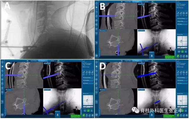
图2. (A)参考架安放固定在锁骨上。(B,C,D)导航系统三维影像引导下,用指示针选择椎弓根最佳的进针点和方向。
1.4导针验证。在椎弓根针道置入导针后,再次O-Arm扫描和成像,冠状位、矢状位和轴位上观察针道的准确性(图3)。
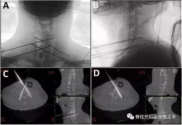
图3. (A,B)椎弓根针道置入导针。(C,D) O-Arm成像后观察针道的准确性。
1.5置钉。用中孔3.5mm丝攻在导针引导下攻丝,选择合适长度的前路椎弓根钉板(Z3,山东威高),在椎弓根钉孔置入长度3.5cm直径4mm的椎弓根钉,在椎体钉孔置入椎体钉(图4)。手术常规冲洗、引流和关闭伤口。手术时间80分钟,其中导航和置钉时间50分钟,出血约80ml。
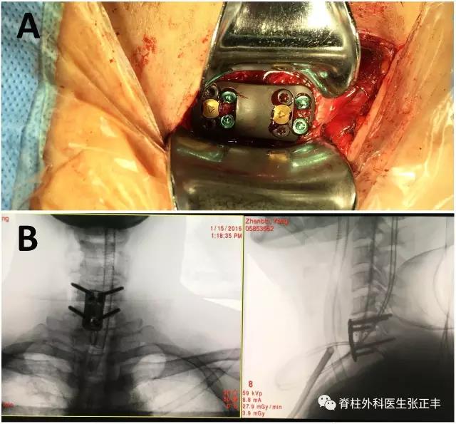
图4. (A)前路椎弓根钉钢板置入后的术中照片。(B)钢板置入后的正侧位片。
1.6术后复查。术后第二天拔出引流管,戴颈托下床活动,左上肢肌力恢复。复查CT显示椎弓根钉位置良好,MRI显示6/7左侧椎间孔减压良好(图5)。
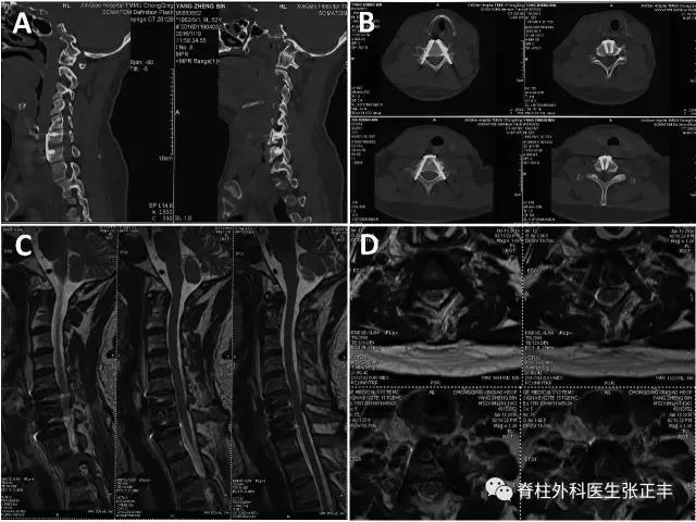
图5.(A,B)CT显示椎弓根钉位置良好。(C,D) MRI显示6/7左侧椎间孔减压良好。
2.讨论
2.1有效性。下颈椎后路椎弓根钉固定一般有三种方法:徒手技术、椎板椎间孔直视技术(laminoforaminotomy)和导航技术。徒手操作和直视技术置钉的准确率报道为72%-99%[12-16],导航技术中,采用骨性标志方法仅有12.5%的准确率,采用术前CT和注册方式可有76%的准确率,而采用术中3D成像和导航可获得81.3%的准确率[17]。采用术中O-Arm 3D图像加导航技术可获得99.2%的有效率,本文采用同样的技术,置入4枚前路椎弓根钉,没有出现置入错误或并发症。作者认为,这种高有效率与术中实时影像和先进的导航器械有关。
2.2安全性。下颈椎后路椎弓根钉技术存在脊髓、神经根椎动脉损伤的风险。在徒手技术中,Abumi等报道在669椎弓根中出现2例神经根痛、1例无神经症状的椎动脉损伤[16];Yukawa等报道419椎弓根置入中出现2例椎动脉损伤和1例神经根痛[13]。在骨性标志方法和术前CT加注册方法的导航技术中,神经血管损伤的发生率可高达37%[17]。而采用术中O-Arm 3D图像加导航技术还没有颈椎神经血管损伤的报道[11],这也是本文采用此技术的原因。
2.3注意事项。由于病例的稀缺,本文仅报道1例术中O-Arm 3D成像和导航下颈椎前路椎弓根钉植入,其有效性和安全性需要更多病例验证。作者报道了采用椎弓根轴位透视法植入CAPS的手术技巧和病例报道[10],由于本文方法病例较少,还无法做统计学比较。为了提高本方法置入的准确性和安全性,作者建议,如果发现置入椎发生可疑位置移动,指示针一定要在骨性标志上验证,甚至重新扫描和注册。
2.4结论。本文首次报道1例采用术中O-Arm 3D成像加导航应用于颈椎小关节骨折脱位前路椎弓根植入,并取得了满意疗效。但由于病例较少,此方法的准确性和安全性需更多病例的验证。
参考文献:
1. Koller H, Hempfing A, Acosta F, Fox M, Scheiter A, Tauber M, et al. Cervicalanterior transpedicular screw fixation. Part I: study on morphologicalfeasibility, indications, and technical prerequisites. Eur Spine J 2008; 17:523–38.
2. Koller H, Acosta F, Tauber M, Fox M, Martin H, Forstner R, et al. Cervicalanterior transpedicular screw fixation (ATPS) – Part II. Accuracy of manualinsertion and pull-out strength ofATPS. Eur Spine J 2008;17:539–55.
3. Koller H, Hitzl W, Acosta F, Tauber M, Zenner J, Resch H, et al. In vitrostudy of accuracy of cervical pedicle screw insertion using an electronicconductivity device (ATPS part III). Eur Spine J 2009;18:1300–13.
4. Koller H, Schmidt R, Mayer M, Hitzl W, Zenner J, Midderhoff S, et al. Thestabilizing potential of anterior, posterior and combined techniques for thereconstruction of a 2-level cervical corpectomy model: biomechanical study andfirst results of ATPS prototyping. Eur Spine J 2010;19:2137–48.
5. Fu M, Lin L, Kong X, Zhao W, Tang L, Li J, et al. Construction and accuracyassessment of patient-specific biocompatible drill template for cervicalanterior transpedicular screw (ATPS) insertion: an in vitro study. PLoS ONE2013; 8:e53580.
6. Chen C, Ruan D, Wu C, Wu W, Sun P, Zhang Y, et al. CT morphometricanalysis to determine the anatomical basis for the use of transpedicular screwsduring reconstruction and fixations of anterior cervical vertebrae. PLoS ONE2013; 8:e81159.
7. Yukawa Y, Kato F, Ito K, Nakashima H, Machino M. Anterior cervical pediclescrew and plate fixation using fluoroscope-assisted pedicle axis view imaging:a preliminary report of a new cervical reconstruction technique. Eur Spine J2009; 18:911–16.
8. Aramomi M, Masaki Y, Koshizuka S, Kadota R, Okawa A, Koda M, et al.Anterior pedicle screw fixation for multilevel cervical corpectomy and spinalfusion. Acta Neurochir (Wien) 2008; 150:575–82 discussion 582.
9. Zhao L, Li G, Liu J, Benedict GM, Ebraheim NA, Ma W, et al. Radiologicalstudies on the best entry point and trajectory of anteriocervical pedicle screwin the lower cervical spine. Eur Spine J 2014;23:2175–81
10.Zhang Z, Mu Z, Zheng W. Anterior pedicle screw and plate fixation for cervicalfacet dislocation: case series and technical note. Spine J. 2016;16(1):123-9
11.Theologis AA, Burch S. Safety and Efficacy of Reconstruction of Complex Cervical Spine Pathology Using PedicleScrews Inserted with Stealth Navigation and 3D Image-Guided (O-Arm)Technology. Spine. 2015;40(18):1397-406
12.Kast E, Mohr K, Richter HP, et al. Complications of transpedicular screwfixation in the cervical spine. Eur Spine J 2006; 15: 327-34.
13.Yukawa Y, Kato F, Yoshihara H, et al. Cervical pedicle screw fixation in100 cases of unstable cervical injuries: pedicle axis views obtained using fluoroscopy.J Neurosurg Spine 2006;5: 488-93 .
14.Yoshimoto H, Sato S, Hyakumachi T, et al. Spinal reconstruction using acervical pedicle screw system. Clin Orthop Relat Res 2005; 431: 111-9.
15.Ludwig SC, Kowalski JM, Edwards CC, et al. Cervical pedicle screws: comparativeaccuracy of two insertion techniques. Spine 2000; 25:2675-81.
16.Abumi K, Shono Y, Ito M, et al. Complications of pedicle screw fixationin reconstructive surgery of the cervical spine. Spine 2000; 25: 962-9
17.Ludwig SC, Kramer DL, Balderston RA, et al. Placement of pedicle screwsin the human cadaveric cervical spine: comparative accuracy of three techniques.Spine 2000; 25: 1655-67
本文来源:脊柱外科医生张正丰


